What is Tendinopathy?

Dr Farrah Jawad
Consultant in Sport, Exercise & Musculoskeletal Medicine
- 29 November, 2018
- Podiatry
- 4 min read
What is Tendinopathy?

Tendons attach muscles to bone, and we have tendons at various places in our bodies. Normally, tendon fibres are linear and organised, like a packet of spaghetti. Sometimes, the tendon fibres can become disorganised, and new blood vessels may form as the tendon tries to heal itself. This can be associated with pain or aching in the tendon, and you may notice swelling over the affected area.
You may see tendinopathy written as tendinitis or tendinosis. Historically, there has been some debate over what is going on in the tendon at a cellular level. That is where this difference in terminology has come from. Understanding what is occurring at a cellular level helps us to understand how tendinopathy occurs and how it is best managed, and in the future, as we understand more about what happens at the cellular level, it may form the basis for new treatments.
What Types of Tendinopathy Are There?
Tendons can be affected very close to the area where they join onto the bone ( insertional tendinopathy) or slightly further from the bone (midportion tendinopathy). Some tendons have a sheath, or covering, around them. Sometimes the tendon sheath can become irritated and inflamed, and we aim on reducing inflammation. The area of the tendon affected can sometimes change the management plan slightly, and focus on reducing inflammation.
What Are The Causes of Tendinopathy?
Tendinopathy can happen for lots of reasons, such as:
- A sudden increase in training load or frequency
- Biomechanical factors
- A group of antibiotics called fluoroquinolones (e.g. ciprofloxacin) can occasionally be associated with tendon problems
- Being older (although tendinopathy can also occur in younger people)
- Some tendinopathies are more strongly associated in certain gender groups; for example, patellar tendinopathy of the knee is more common in males. This may be due to certain biomechanical factors, different sporting behaviours and hormonal factors.
- Being overweight
There is evidence emerging about the genetic factors involved in tendinopathy.
What Are The Symptoms Of Tendinopathy?
-
Symptoms of tendinopathy can vary in intensity and onset, but they often develop gradually and may worsen with continued use of the affected tendon. Common signs include:
-
Aching, discomfort, or pain and tenderness located over the injured tendon. This pain may be mild at first, appearing only during or after activity, but it can progress to persistent discomfort even at rest if the condition worsens.
-
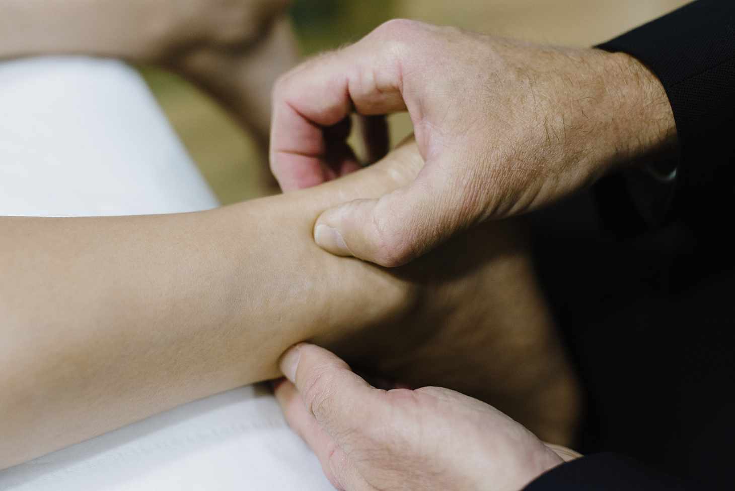
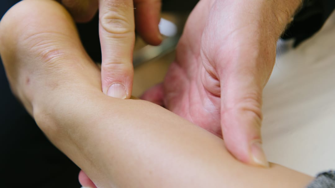
Tenderness to touch over the affected area, which may make pressing or squeezing the tendon uncomfortable.
-
Swelling or a noticeable lump that can sometimes be felt or seen along the tendon. This swelling may be due to inflammation, fluid buildup, or thickening of the tendon tissue.
-
A feeling of stiffness in or around the tendon, especially after periods of inactivity such as sleeping or sitting for a long time. This stiffness often eases with gentle movement but may return after overuse.
In some cases, individuals may also notice reduced strength or mobility in the nearby joint, or a creaking or grating sensation (known as crepitus) when the tendon is moved. These symptoms can impact daily activities, sports participation, and overall function if not addressed promptly.
-

Diagnosis and Investigations
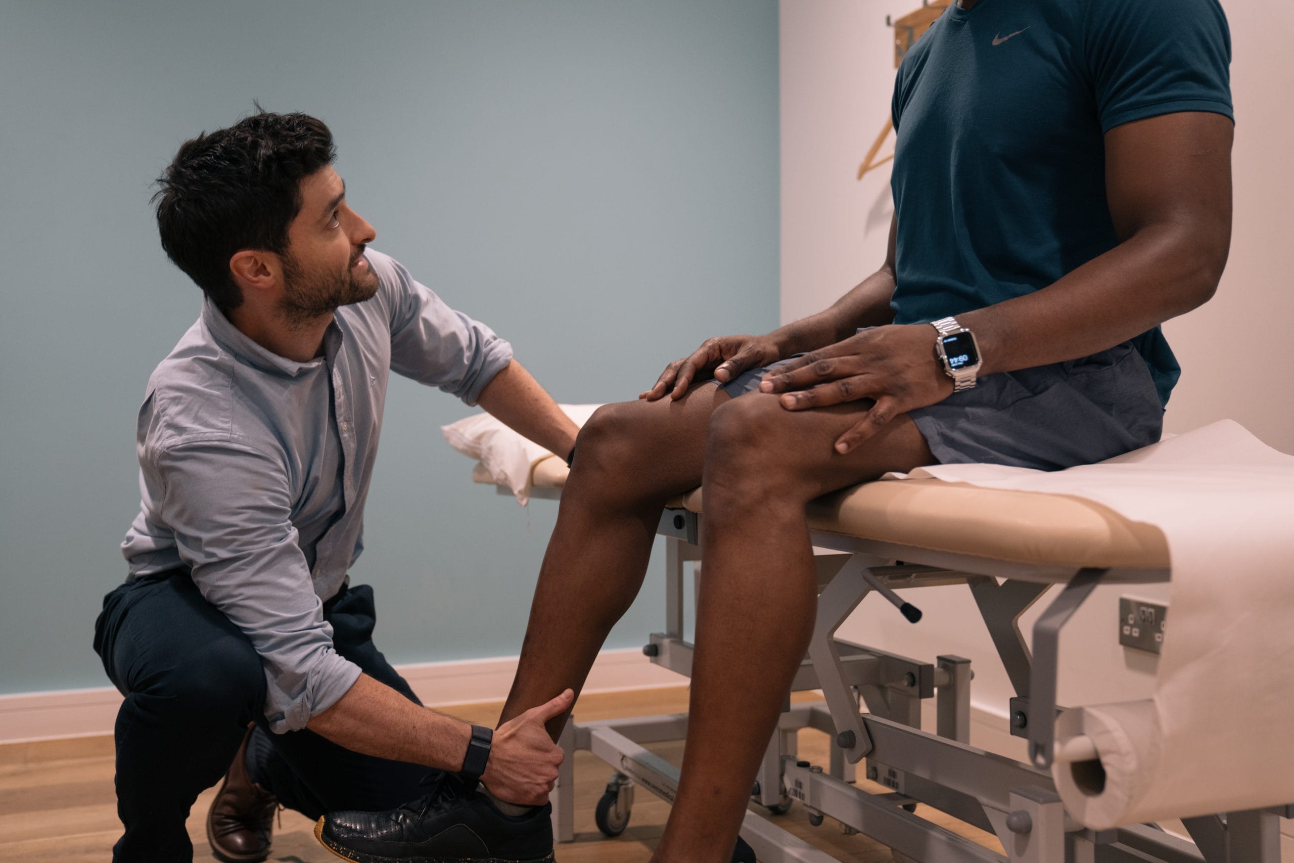
Usually, people will describe a history of symptoms which will make the Sports Doctor consider tendinopathy as the diagnosis, and there can be signs of this that they will see when they examine you. The doctor will ask questions about your past medical history and any other symptoms you may have.
Many tendons can be well visualised with an ultrasound scan, which a Sports Doctor can perform for you. Ultrasound scans are relatively quick, painless, involve no radiation and can allow a dynamic assessment of the tendon. If there is tendinopathy, this will be seen on the scan.
Tendons can also be seen using an MRI scan. If you require a scan, your sports doctor will be able to discuss with you which type of scan is most appropriate for you based on their assessment.
Management Strategies For Tendinopathy
- Pain relief can be achieved by avoiding aggravating activity, offloading the affected area, and applying an ice pack. A Sports Doctor may suggest a non-steroidal anti-inflammatory medication (NSAID), which may be helpful initially
- Modifying activity, such as by changing your training regime
- Assessment of biomechanics – a therapist may have some suggested targets for rehabilitation, which may be beneficial. A therapist may suggest equipment or footwear modification, for example
- Seeing a podiatrist can be helpful for lower limb issues
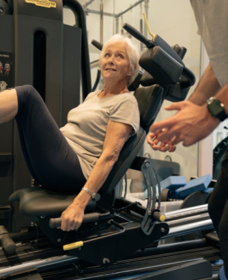
- A physiotherapist can help guide you through an exercise programme
Most people will get better with the above measures to reduce pain. It can take time and commitment to an exercise programme to see improvements. Your physiotherapist will give you exercises to do to work on between physiotherapy sessions. As these become easier, they will modify the exercises to help the tendon get back to functioning normally.
We know that how the tendon appears on a scan does not correlate well with a person’s symptoms. Furthermore, tendon appearances on scans can lag behind a patient’s improvement in their symptoms of tendonitis. Therefore, doctors don’t use scans to check if the tendon appears more “normal” or not during a treatment programme, except for research purposes.
Prevention
Clearly, some of the contributing factors to developing tendinopathy are modifiable (e.g. training load and biomechanics) and some are not (e.g. normal ageing). Various members of the team at Pure Sports Medicine – such as a Sport, Exercise & Musculoskeletal Consultant, Physiotherapist, Strength and Conditioning Coach, Podiatrist – can help to reduce your chances of being affected by tendinopathy. A Sports Doctor can make the diagnosis for you initially and refer you onto other members of the team as part of a comprehensive management strategy.
If you are interested in finding out more about services offered at Pure Sports Medicine, find out more or book an appointment.

Advice
Over the last 20+ years our experts have helped more than 100,000 patients, but we don’t stop there. We also like to share our knowledge and insight to help people lead healthier lives, and here you will find our extensive library of advice on a variety of topics to help you do the same.
OUR ADVICE HUBS See all Advice Hubs

